
Introduction
Changes in breathing patterns have been known to be associated with respiratory and non-respiratory diseases for many years [1–4]. Monitoring changes in breathing patterns over time could therefore provide useful information about respiratory health status or be an outcome measure for interventions. Before any parameter can be useful as a clinical monitoring tool or outcome measure, there is a need to assess its stability within individuals [5].
Breathing pattern is traditionally assessed using observation and manual palpation. The only element of breathing pattern to be routinely quantified during spontaneously breathing is respiratory rate [6]. However, breathing pattern is much more complex involving components of timing, volume, movement of the chest wall, and rhythmicity [7–9]. Capturing and quantifying all these elements within routine clinical practice is challenging, but there are several technologies available to quantify the parameters of breathing within the laboratory setting. A pneumotachograph is considered to be the ‘gold standard’ for recording air flow and capturing parameters of timing and volume but involves mouthpieces and facemasks that can alter the pattern of breathing [10, 11]. Respiratory inductive plethysmography (RIP) has been validated as the method of choice for recording breathing parameters non-invasively; it evaluates respiratory parameters, including timing, volume, and the involvement of the rib cage and abdomen in tidal volume, by measuring the extent of movement in the rib cage and abdominal areas during inhalation and exhalation [12].
The stability of some timing and volume elements of breathing pattern within individuals has been previously studied, albeit infrequently [13]. Shea et al. [14, 15] conducted studies using RIP to assess the stability of breathing pattern parameters in healthy adults during wakefulness and sleep. They reported a high degree of stability of breathing pattern within individuals, with respiratory rate being the most reproducible variable. Benchetrit et al. [13] used a different measurement technique (pneumotachograph), but also found the timing and volume parameters of the breathing pattern remained consistent over time. Recently, Hedge et al. [16] used RIP to record timing, volume, and thoracoabdominal motion in healthy adults during three level of step exercise, finding a high degree of stability in breathing patterns throughout the activity.
Although there is some evidence that the timing and volume elements of breathing pattern remain stable over time within individuals, there is little information about the relative contributions of the ribcage and abdomen to breathing, or the impact of changing body position on the stability of these parameters as it is evidence that posture has an impact on chest wall motion. Ribcage and abdominal movements are often of particular interest to physiotherapists, who aim to manipulate them as part of breathing retraining programmes [17, 18]. This is the first study to examine the stability of the relative contributions of the ribcage and abdomen to breathing in two positions, by recording breathing pattern using RIP in healthy adults with in the sitting and supine positions
Subjects and methods
Design
An observational single group repeated measures design was used to examine the stability of breathing pattern components: (1) within recording sessions, (2) between recording sessions on a single day, and (3) between recording sessions on different days.
Participants
Healthy adults aged 18 years or older who were able to provide informed consent were eligible for inclusion in the study. Anyone with a history of respiratory disease was excluded.
Sample size
The sample size was determined using a reliability sample size calculator [19] within Microsoft Excel. The calculation considered four key factors: the number of repeated measures (i.e., the number of measurements taken), the intraclass correlation coefficient (ICC) based on pilot study data from 20 participants, a significance level of 5%, and a 95% confidence interval (CI) width of 0.5 for TE and 0.3 for other components. Appendix A provides an example of the Excel spreadsheet used for the sample size calculation.
As shown in Table 1, the maximum required number of participants was 51, leading to the decision to recruit 51 participants. The largest prior study investigating the reliability of breathing patterns in adults involved 41 participants, making a sample size of 51 appropriate for this research.
Recruitment
Recruitment was carried out at the University of Southampton by placing posters around the campus. Anyone interested in taking part was advised to contact the researcher directly via telephone or email. Upon contact, the researcher checked whether the volunteer met the study eligibility criteria and subsequently made a convenient appointment for the baseline research appointment.
Data collection procedure
Participants attended four data collection sessions over two separate days (4–6 days apart depending on availability) at the Faculty of Health Sciences, University of Southampton. At the first session on day 1, after giving written informed consent, demographic data (age and sex), anthropometric data (height and weight) and data relating to general health were recorded.
Breathing pattern data were then recorded using RIP. An Inductotrace system (Respitrace®, Ambulatory Monitoring Inc. New York) was used to record respiratory parameters relating to the regional contributions of the ribcage and abdomen. Participants were fitted with two elasticised belts (Inductobands) embedded with Teflon insulated wires. These were fastened around the bare chest (females retained their undergarments) at the level of the ribcage (just below the axillae) and the abdomen (just below the lowest vertebral ribs). A custom-built analogue-to-digital converter was used to convert the signals acquired by the RIP to digital form with a sampling frequency of 10 kHz. Calibration of the RIP system was performed using a fixed calibration procedure previously described by Banzett et al. [20] to account for relative differences in the relationship between movement and respiratory volume in the ribcage and abdomen. Respiratory parameters were extrapolated from the recorded displacement of the ribcage and abdomen during inspiration and expiration.
Breathing pattern recording procedure
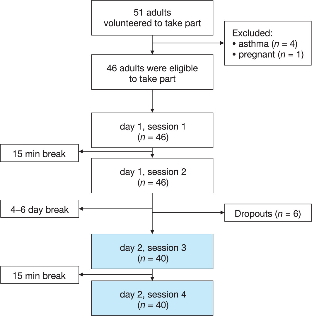
The same recording procedure was used at each data collection session. Breathing patterns were first recorded in sitting, and then in the supine position, for 15 min each. Figure 1 illustrates the sequence and timeframe of each breathing pattern recording, and the number of participants who attended each data collection session.
Breathing parameter extraction
Breathing parameters were derived from the RIP signal data using a customised peak detection algorithm developed in Matlab (2009)® by a postdoctoral research fellow from the Institute of Sound and Vibration Research, University of Southampton). Breathing parameters were calculated by identifying the peaks and troughs (defined as the lowest and highest points respectively) of each individual displacement signal recorded from the two bands of the RIP, and from their sum. The regional contributions of the ribcage and abdomen during inspiration and expiration were defined as the amplitude change in the signal from the ribcage or abdomen band relative to the amplitude of the sum of the two signals, and expressed as a percentage. Other parameters extracted were: (1) inspiration and expiration time (TI and TE) in seconds, defined as the time from a trough to the next peak, and time from a peak to the next trough respectively, (2) inspiration and expiration volume in arbitrary units, defined as the amplitude change from trough to peak, and from peak to trough respectively. The mean of each parameter over each recording period was then calculated. Respiratory rate in breaths per minute was also calculated by summing the TI and TE for each cycle to determine the total breathing cycle duration and taking the reciprocal of the mean of this measure over the 15 min recording period.
Statistical analysis
The mean and standard deviation of each of the eight extracted breathing parameters were calculated for each 15 min recording period. Estimates of absolute and relative reliability were calculated for only four breathing parameters because some of the eight extracted parameters were interrelated, or were reciprocals of each other (such as percentage of ribcage and abdominal contributions to total movement). The representative parameters selected for reliability analyses were inspiration and expiration time, respiratory rate, and the contribution of the ribcage to expiration. Absolute and relative reliability in the sitting and supine position were assessed: (1) within a single recording session, (2) between recording sessions on a single day, and (3) between recording sessions on two different days. For the within sessions analyses, the data from each session were divided into five sequential segments of three minutes each, and the second and fourth segment of each 15 min period were compared. The decision to ignore the first segment was taken in order to enable the participants to settle, while the fifth segment was ignored because it was observed that participants became more restless towards the end of the 15 min recording. The between session analyses were performed using data from sessions one and two (on day one), and between sessions three and four (on day two). The between day analyses were performed using data from session two (day one) and session four (day two). This decision was made because it was considered that breathing patterns obtained during the second session of each day may have been more ‘natural’ since participants were more accustomed to the data collection procedure. One-way repeated measures analysis of variance (ANOVA) tests were used to generate the within subjects standard deviation (WSSD). The WSSD was used to assess the absolute reliability of each selected breathing pattern component. The WSSD is calculated as the root square of the within subjects mean square (derived from the ANOVA table), to indicate the level of uncertainty that occurs within a measurement. Smaller WSSD values provide an indication that measurements are more reliable [21]. Although hypothesis testing is not the primary aim of reliability studies, the ANOVA tests allowed us to look for any statistically significant differences in the means of each selected breathing parameter within and across data collection sessions. The intra class correlation coefficient (ICC) was calculated to assess the relative reliability of breathing pattern components at different time points. Bland and Altman 95% limits of agreement (LA) were calculated to assess the level of agreement between measurements within a single session, for both the sitting and supine data [22]. This method was also used to compare data between sessions in one day and between day one (session two) and day two (session four). Smallest real difference (SRD) calculations were made to determine how much change needs to occur between measurements to be considered a ‘real’ change, as opposed to measurement error. The SRD calculation has previously been used as a measure of responsiveness to change [23].
Search strategy
The following electronic databases were used: CINAHL Plus with full text, MEDLINE, Cochrane Central Register of Controlled Trials (CENTRAL). Key search terms: breathing pattern components, breathing pattern test re-test reliability, breathing pattern at rest, thoraco-abdominal motion, respiratory inductive plethysmography (RIP), and validity of RIP. Also, a manual search of respiratory journals was conducted, including: Chest, Respiration Physiology, Journal of Applied Physiology, Respiration, American Journal of Respiratory and Critical Care Medicine.
Results
Fifty-one adults (12 males) initially volunteered. Five were excluded (four reported that they had asthma and one was heavily pregnant). Forty-six participants completed data collection on day 1 (session 1 and 2), but six subsequently dropped out citing ’limited time capacity’ as the primary reason for dropout. Forty participants generated complete data sets from all sessions, but technical problems with the RIP resulted in corrupted data from two participants, hence their data sets were subsequently removed. Data presented in this paper are therefore represent the 38 participants (six males) who completed all four data collection sessions and generated usable data. Demographic and anthropometric data for these 38 have been presented in Table 2. The mean age for the whole group was 31 years (SD =7), with a mean body mass index (BMI) of 25 (SD = 5).
Table 2
Demographic and anthropometric characteristics according to gender for participants who attended all four sessions (n = 38)
Breathing parameters
Tables 3–6 provides a summary of the mean breathing parameter data obtained for all participant over the four sessions in the sitting and supine positions. These data have been presented for each recording session in sitting and supine (Tables 3 and 4), and within recording sessions (Tables 5 and 6). Within session analyses were performed for all four sessions, they all generated similar results and so only within session 2 and 4 are presented here. Between session and between day analyses were also carried out for every possible combination presented in Tables 7 and 8.
Table 3
Breathing parameters in the sitting position during each session: descriptive statistics
[i] TI – inspiratory time, TE – expiratory time, BC – breathing cycle duration, RR – respiratory rate, %RCInsp – ribcage contribution to tidal volume during inspiratory phase, %RCInsp – ribcage contribution to tidal volume during inspiratory phase, %RCExp – abdominal contribution to tidal volume during expiratory phase, %ABExp – abdominal contribution to tidal volume during expiratory phase
Table 4
Breathing parameters in the supine position during each session: descriptive statistics
[i] TI – inspiratory time, TE – expiratory time, BC – breathing cycle duration, RR – respiratory rate, %RCInsp – ribcage contribution to tidal volume during inspiratory phase, %RCInsp – ribcage contribution to tidal volume during inspiratory phase, %RCExp – abdominal contribution to tidal volume during expiratory phase, %ABExp – abdominal contribution to tidal volume during expiratory phase
Table 5
Within session descriptive statistics for all breathing parameters in the SITTING position
[i] TI – inspiratory time, TE – expiratory time, BC – breathing cycle duration, RR – respiratory rate, %RCInsp – ribcage contribution to tidal volume during inspiratory phase, %RCInsp – ribcage contribution to tidal volume during inspiratory phase, %RCExp – abdominal contribution to tidal volume during expiratory phase, %ABExp – abdominal contribution to tidal volume during expiratory phase
Table 6
Within session descriptive statistics for all breathing parameters in the SUPINE position
[i] TI – inspiratory time, TE – expiratory time, BC – breathing cycle duration, RR – respiratory rate, %RCInsp – ribcage contribution to tidal volume during inspiratory phase, %RCInsp – ribcage contribution to tidal volume during inspiratory phase, %RCExp – abdominal contribution to tidal volume during expiratory phase, %ABExp – abdominal contribution to tidal volume during expiratory phase
Table 7
Reliability estimates of breathing parameters in the SITTING position (n = 38)
Table 8
Reliability estimates of breathing parameters in the SUPINE position
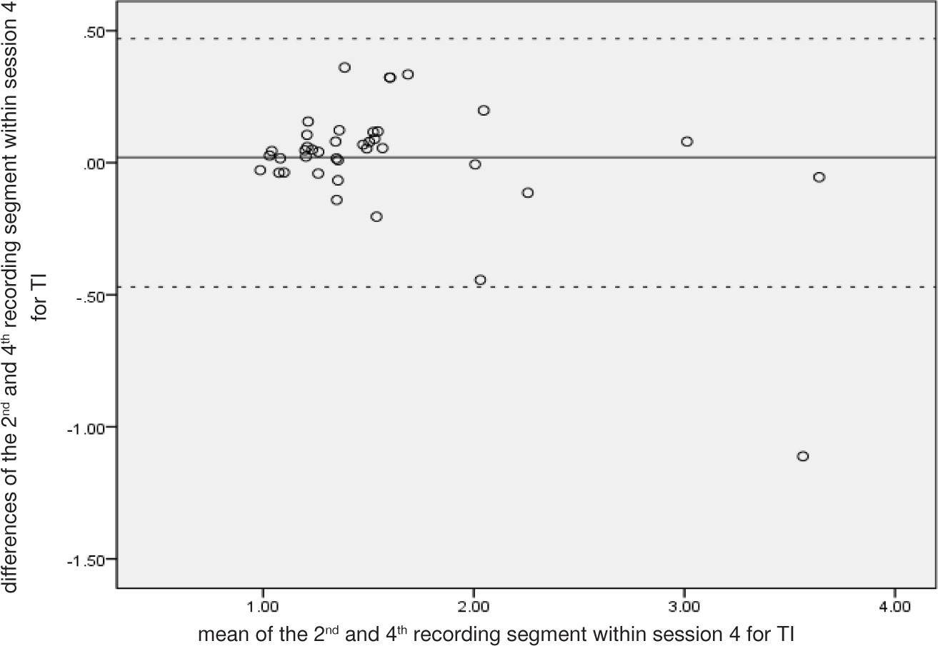
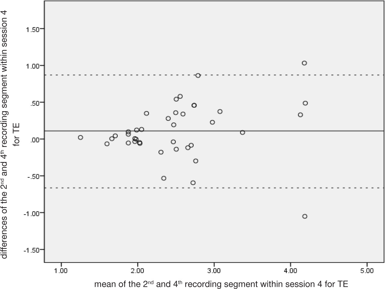
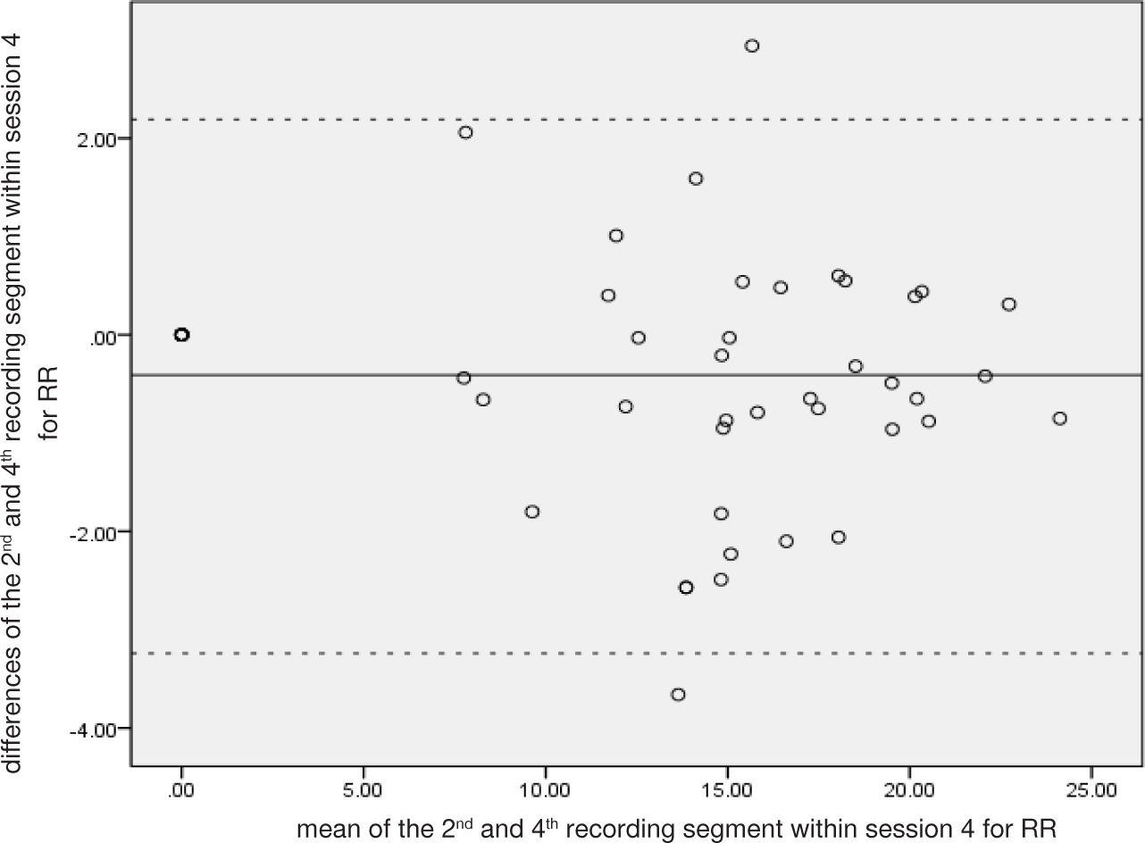
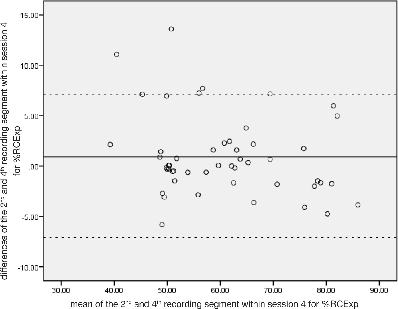
In both sitting and supine positions, the ANOVA indicated no statistically significant differences in cohort mean values in nearly all parameters across all comparisons (p < 0.05). The exceptions were respiratory rate within session 4 (sitting) (p = 0.009) and inspiratory time between sessions 3 and 4 (supine) (p = 0.02), but neither were associated with clinically meaningful differences. ICC values were uniformly high (> 0.7) in data from both positions and across all comparisons, suggesting good relative reliability. The WSSD values were used to calculate the smallest real difference (see Tables 7 and 8). The Bland and Altman analysis (the calculation of 95% limits of agreement and the generation of Bland and Altman plots) indicated no bias between measurements, as there is no evidence of funnelling and there is a consistent level of variation indicating good agreement across all breathing parameters in both positions. Figures 2–5 illustrate the Bland–Altman plots for breathing pattern components for within session 4 in sitting position.
Discussion
Our main findings suggest that breathing pattern parameters remain stable within individuals over time in both sitting and supine positions, with the sitting position showing slightly higher levels of stability. The novelty of our work comes from the analysis of the respective ribcage and abdominal contributions to respiratory movement. In addition to the timing and volume parameters, these movement parameters also show remarkable stability over time. Although it is known that there is considerable variability breath to breath within individuals in all elements of breathing pattern [7, 12, 14], it seems that summary data from breaths recorded over periods from three to 15 min remain consistent. There are insufficient data related to many of the extracted parameters in the public domain to know if any of the small measurable differences have any clinical relevance. The cohort mean differences between sessions and between days for all components were all smaller than the SRD values derived from within session data, however, suggesting no ‘real’ differences between recordings.
There has been little published research in this area, but our findings on timing and volume parameters support those of previous authors. Shea et al. [14, 15] conducted studies of breathing patterns in both awake and sleeping healthy adults in supine. In their awake study, the breathing patterns of 41 healthy adults aged between 19 and 32 were recorded four times for five minutes each over the course of two days using RIP. Tidal volume, inspiration and expiration time, respiratory rate and the proportion of time spent on inspiration (expressed as a percentage) were examined. They reported high degree of reproducibility of breathing pattern within individuals, with respiratory rate; being the most reproducible parameter. Similar findings were reported for their sleep study. Benchetrit et al. [13] used a pneumotachograph to assess stability of elements of breathing pattern over a longer period of time. Breathing patterns were recorded twice over five years, the analysis was conducted using 50 breaths per participant, with any sighs or pauses manually excluded from the recordings. The results indicated that variations within individuals were significantly smaller than those observed between individuals, even when some participants experienced changes in circumstances such as weight gain, respiratory illnesses, or learning to play wind instruments. Furthermore, Hedge et al. [16] used RIP to examine the stability of timing, volume, and thoracoabdominal motion in 10 healthy adults during physical exercise. The results suggest that the components of the breathing parameters remained stable throughout the 3 level of step exercise. However, no base line data was collected. It therefore seems that irrespective of the measurement tool used to record breathing patterns, the elements of respiratory timing and volume remain stable within healthy individuals. Although supine is often the preferred position in research studies, our findings suggest that recording breathing patterns in sitting is equally useful, as they show similar or higher levels of stability for all parameters. visual inspection of the our data shown higher WSSD values and the wider 95% LA in the supine position in comparison to the sitting position, indicating more variability and lower agreement in the supine position for all comparisons. Moreover, in general, the standard deviations within the descriptive data were greater for the supine position, than the sitting position, again indicating more variability of breathing pattern components when measured in the supine position.
Our novel findings concerning the stability of ribcage and abdominal contributions to respiratory movement cannot be compared to previous literature. Some of our descriptive data for these parameters are, however, similar to others. Tobin et al. [1] reported RIP data from 47 healthy adults of similar ages to our sample and found the average ribcage contribution in supine to be 42% (it was 36–41% in our supine data). Parreira et al. [24] also used RIP and reported ribcage percentage contributions to be between 40 and 46% in healthy female adults. Mendes et al. used Opto-Electronic Plethysmography (OEP) and reported ribcage contributions of 31% and 35% for male and female respectively [2]. These findings indicate that chest wall contributions found in this study are similar to previous studies, and despite acknowledged gender and posture differences, seem to remain consistent even when different measurement tools are used.
Breathing pattern is complex and as Benchetrit [7] states, there are ‘an infinite number of possible combinations of ventilatory components and airflow shape capable of achieving the same minute ventilation’. What is remarkable is that individuals appear to select one particular pattern from within this complexity, and that this selection appears to be a relatively stable characteristic over time within adults Fadel et al. [12] examined breathing patterns in 20 adult healthy participants on one occasion, using fractal analysis. They report detectable stable patterns within the seemingly random breath to breath variability of rate, timing and volume parameters.
Our research suggests that breathing pattern parameters also remain stable within individuals over time. This is useful information for those physiotherapists who use interventions like breathing retraining. Breathing retraining for asthma aims to ‘normalise’ various aspects of respiratory pattern in terms of timing (slowing rate), volume (reducing depth) and use of abdominal movement over use of ribcage for expansion [8, 25, 26]. At present, however, there is no objective evidence that these goals are achieved. Having established that these parameters are stable within healthy individuals, it becomes possible to consider their use as objective measures to assess the effects of such interventions.
Limitations
The use of convenience sampling from the available adult population in and around a university campus may have led to an unrepresentative sample, as in this study we recruited 32 females and only 6 men. The lack of significant differences seen in the ANOVA calculations may therefore have been due to insufficient statistical power to detect a difference. Sample size has the opposite effect on ICC, however, as a small sample could potentially overestimate variance, and would therefore be less likely to generate results that support good reliability. Recording ‘normal’ breathing pattern is inevitably challenging because informed consent means that people are aware that their breathing is being studied, and this awareness plus the instrumentation are likely to affect breathing pattern.
Conclusions
This is the first study to report the stability of ribcage and abdominal contributions to respiration within healthy adults over time at rest. Using data averaged over a 15-minute period, breathing patterns of healthy adults remained stable within sessions, between sessions and between days in terms of timing and volume components, and ribcage/abdomen contributions to respiratory movement. These findings, taken together with existing evidence of the individuality of breathing pattern, have relevance for physiotherapists aiming to alter any of the elements of breathing pattern during their therapeutic interventions.