Introduction
Low back pain (LBP) refers to the pain felt between the lower border of the ribs and the intergluteal cleft [1]. LBP in itself is not a disease but a symptom whose origin is often difficult to determine [1].
Even though LBP problems affect a wide range of populations of all ages, from teenagers to the elderly, the condition clearly becomes more common with age [1–4]. It is believed that the majority of the adult population will experience LBP at a certain stage in their lives [2, 4]. LBP is also considered the most common cause of disability [1–3]. The condition is as common in highly developed countries as it is in developing or underdeveloped ones. Women suffer from the condition more frequently and thus are more likely to seek medical help to improve their health [1–4].
In most patients, it seems impossible to determine the specific cause of LBP [2–4]. Imaging studies such as magnetic resonance imaging (MRI) or computed tomography (CT) performed on patients with LBP often display various anatomical abnormalities, but the same defects are also often found in patients who do not report pain [1]. In young patients, the presence of certain abnormalities on imaging studies may increase the chance of LBP occurring in the future [1].
It is assumed that LBP may be caused by a combination of several factors, including biophysical features (muscle mass, composition or volume, anatomical structure), psychological factors (fear, depression) and social aspects (level of education, access to health care, quality of health care, occupation, financial situation, etc.) [2]. These complaints are sometimes caused by specific conditions, such as vertebral fractures, neoplastic lesions, or inflammatory diseases, but these require specific treatment and constitute a small percentage of LBP cases [2–4].
A method that is gaining popularity as a treatment option for LBP is dry needling (DN). The treatment involves identifying and then puncturing active myofascial trigger points (MTrPs) in skeletal muscles. This causes a local twitch response (LTR) in the tense muscle band, resulting in its relaxation [5]. A puncture is done with a thin acupuncture needle, but the method is strictly associated with conventional Western medicine [6]. DN is a minimally invasive method, and it is also simple to perform [5]. It can be effective in providing pain relief, and it improves the functional status of the patient [5, 7]. Despite the potential benefits of DN in the treatment of LBP, the number of research studies and publications on the therapy is low, and the available articles describing positive effects of the treatment emphasise the need of further research into the effectiveness of DN in LBP [7, 8].
The purpose of the present study is to investigate the effectiveness of dry needling (DN) in the context of treating LBP-related symptoms over a period of four weeks following laparoscopic removal of an ovarian cyst in a female patient.
Subjects and methods
The patient is a female aged 24, with a self-reported height of 182 cm and a body weight of 72 kg (BMI 21.74 kg/m2– within normal limits). The average level of physical activity per week is, according to the patient’s declaration, 2 hours. The patient has no additional comorbidities or addictions. She underwent a laparoscopic surgery to remove an ovarian cyst 2 years ago. She reports back pain in the lumbar area, which persists at an average of 5 points on the Numeric Rating Scale (NRS) scale and started approximately 4 years ago. The patient was not taking any painkiller medication during the course of this study.
The inclusion criteria were the following: age 20 to 30 years, low back pain rated at 4 to 9 on the NRS persisting for at least six weeks, and a history of abdominal surgery a minimum of two years ago.
The exclusion criteria were a lack of consent to participate in the study and contradictions to the dry needling procedure.
The project was designed as an observer-blinded trial, where blinding was achieved by separating the study from the therapy. The researchers did not know whether or what kind of therapy the patient was undergoing; the person performing the therapy, in turn, did not know the results of the patient’s subsequent tests.
Measuring tools
The patient’s functional status was assessed using the Oswestry Disability Index (ODI). This questionnaire is divided into ten categories, including pain severity, pain while lifting, sitting, sleeping, travelling, daily care, walking, standing, socialising, as well as the assessment of changes in pain severity. Each category is scored from 0 to 5 points, giving a maximum of 50 points. The score ranges for particular results are established as follows: 0 to 4 points – no disability, 5 to 14 points – mild disability, 15 to 24 points – moderate disability, 25 to 34 points – severe disability, 35 to 50 points – total disability [9].
In order to assess the patient’s pain level, a NRS of 0 to 10 was selected, where 0 means the absence of pain, and 10 means the highest imaginable pain level [10].
The ranges of motion of the lumbar spine in the sagittal and frontal planes were measured using a Saunders digital inclinometer (Baseline Digital Inclinometer, The Saunders Group Inc, Chaska, MN, USA) [11]. The Y-Balance test was used to assess functional capacities in the anterior, postero-lateral, and posteromedial planes for the patient’s lower body [12].
In addition, the viscoelastic properties of soft tissues in the lumbar and hip sections were assessed. The points at which the measurements were made were the so-called coordination centres and were taken from the concept of Fascial Manipulation®. These were the points: AN and RE Lu and Cx (both sides) anatomically. It was 8 points in total (4 for the Lu segment and 4 for the Cx segment). These points, which lie on the rectus abdominis muscle (AN Lu) (lateral to the navel) and on the erector spinae muscle (RE Lu) (at the level of L1) and below the inguinal ligament (above the iliolumbar muscle) and on the gluteus maximus muscle (above the ischial tuberosity, were evaluated for movements in the sagittal plane using myotonometry (MyotonPRO Tallinn, Estonia). The soft tissue properties measured included frequency (F), dynamic stiffness (S), decrement (D), time required for regaining tissue relaxation (R) and creep (C) [13–15]. The parameters were also evaluated in the area of abdominal scar tissue resulting from the surgical procedure.
Experiment protocol
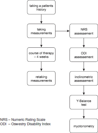
Measurement procedures were carried out in the following sequence: assessment of pain levels using the NRS, assessment of functional status using the ODI questionnaire, evaluation of mobility ranges in the sagittal and frontal planes using an inclinometer, assessment of functional capacity in the anterior, posterolateral and posteromedial planes for the lower body using the Y-Balance test, and finally myotonometry.
As for inclinometric measurement, the result is the arithmetic average of three subsequent measurements of one movement in a given plane. In the case of the Y-Balance test, prior to the measurement, the patient performed six ‘warm-up’ repetitions for each plane, for each lower limb tested, in order to get familiar with the test being performed. Measurements were performed using an original Y-Balance test kit, and the patient was not wearing shoes at the time of testing. The result of the Y-Balance test is the arithmetic average of three measurements taken in each plane for each lower extremity.
Measurement procedures were carried out immediately before the first therapy session, and the post-test was conducted immediately after the last therapy session. Both measurements were performed by the same person at similar times of the day and under similar conditions.
Therapeutic procedures
DN therapy was performed using needles by yellowSPORT (Katowice, Poland) made of 0.3-mm-thick surgical steel. The length of the needle to be used was selected based on the puncture site.
The muscles punctured as part of the therapy included the erector spinae, gluteus maximus, gluteus medius, and piriformis [7–8].
Prior to the application of the needles, the therapist disinfected their hands, put on disposable gloves and disinfected the puncture site. The therapist was unaware of the results of the patient’s functional test (observer-blinded trial).
The testing protocol was the following:
Erector spinae – after palpation of the spinous processes of the lumbar vertebrae, the insertions were performed along the fibres of the spinal erectors at the level of spinal segments L1 to L5; each needle was positioned caudomedially with an inclination of approximately 30°. The technique of stimulation was rhythmic ‘pistoning’ of the needle. After the insertion of the needles, the application was left undisturbed for 15 min, and then the muscle was stimulated again by ‘pistoning’ until the insertion was completed [7, 8]. The needle used was 25 mm long.
Gluteus medius and gluteus maximus muscles – after palpation of the iliac crest and tight bands within the bulk of the muscles, the insertion occurred perpendicularly to the skin surface in the region of increased tension. A stimulation technique of rhythmic ‘pistoning’ of the needle was utilised until LTR was achieved. After the needle’s insertion, the application was left undisturbed for 15 minutes, and then the muscle was again stimulated by ‘pistoning’ the needle until the insertion was completed [7, 8]. The needle used was 75 mm long.
Piriformis muscles – after palpation of the lateral border of the sacrum, the greater trochanter of the femur and tight bands within the bulk of the muscles, the insertion was applied perpendicularly to the skin surface in the region of increased tension. A stimulation technique of rhythmic ‘pistoning’ of the needle was utilised until LTR was achieved. After the needle’s insertion, the application was left undisturbed for 15 min, and then the muscle was again stimulated by ‘pistoning’ until the insertion was completed [7, 8]. The needle used was 75 mm long.
The therapy was carried out twice a week over a period of four weeks, with a minimum of two days’ breaks between therapy sessions in any given week [7, 8].
Results
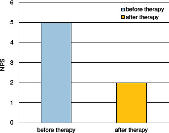
The patient’s reported pain level decreased after the intervention; prior to the treatment, the patient rated the pain severity at 5 points on the NRS, whereas after the therapy, the reported pain level was 2 points on the NRS. Figure 2 graphically demonstrates the result.
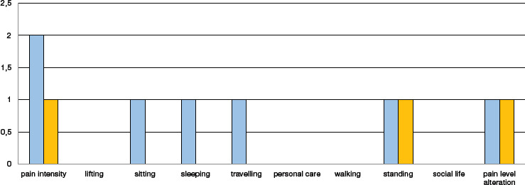
The patient’s functional status, assessed according to the ODI questionnaire, improved. Before the intervention, it was rated by the patient at 7 points. After the therapy, the score according to the ODI questionnaire was 3. Figure 3 graphically demonstrates the result, along with particular categories of the ODI questionnaire.
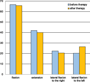
In the inclinometric test, there were no apparent changes for flexion, extension, and lateral flexion to the right. A significant change in the result was noted for lateral flexion to the left; the improvement in the averaged result exceeded 6 degrees. Figure 4 shows the averaged results of all the measurements. The numbers are presented in degrees.
Assessment of the right lower limb functional capacity for the frontal, posteromedial, and posterolateral planes with the use of the Y-Balance test also showed no obvious changes before and after the intervention. A significant change in the result was only noted for the posterolateral plane of the left lower limb: the improvement in the averaged result exceeded 16 cm. The results of particular measurements for the right and left lower extremities are presented in Tables 1 and 2, respectively. The results are presented in centimetres.
Table 1
Y-Balance test results for the right lower limb
Table 2
Y-Balance test results for the left lower limb
In the myotonometric test, the biggest differences are noted in the dynamic stiffness parameter (S) over almost the entire targeted area. A similar dependence can be observed in the parameters of relaxation (R) and creep (C). It is also worth noting the increased tonic frequency in the tissue (F) that results from the intervention. The results of the myotono-metric study are presented in Tables 3 and 4.
Table 3
Results of myotonometry – lumbar spine movements in the sagittal plane
Table 4
Results of myotonometry in the area of postsurgical scar tissue
Discussion
The results obtained in the present study suggest that therapeutic intervention of dry needling carried out in the course of LBP, in the form described in the article, can be an effective method of relieving low back pain. The results, as based on the patient’s perceived pain experience (NRS and ODI questionnaire), indicate a significant improvement in this regard. Within the framework of the proposed methodology, no improvement was observed in the patient’s functional capacity. The results obtained from myotonometric measurements suggest that the intervention through dry needling leads to soft tissues being toned and symmetrised.
The results indicative of pain reduction and tension symmetrisation within soft tissues are an excellent starting point for functional improvement in the patient. Perhaps such an effect might also have been achieved, but the very short time between the end of the therapeutic intervention and the repeat examination (measurement taken immediately after the end of the treatment) did not allow us to demonstrate such a correlation.
A study by Loizidis et al. [16] showed similar effects of dry needling interventions in patients with LBP. Patient-reported pain levels decreased markedly, and functional improvement was also noted in the patients undergoing the intervention.
DN therapy is also effective when combined with other physiotherapy treatments. Pain reduction in sciatic hyper-algesia has been observed after DN therapy combined with neurodynamic treatments [17].
As noted in the introduction, LBP refers to a wide range of conditions, and pain relief achieved through dry needling alone may not be sufficient in the overall treatment process. Medical functional training seems to be an appropriate complement to the proposed therapeutic intervention [18] as it clearly improves the patient’s functional parameters. The use of dry needling as a means of reducing pain and normalising tissue properties could constitute a good starting point for the consolidation and progression of the achieved effects by implementing medical training.
Such conclusions have been drawn from research conducted by Martín-Corrales et al. [19]. According to this study, complementing a medical training program with dry needling may result in significantly better patient pain management compared to a control group where dry needling was not introduced as part of the therapy.
Similar results regarding the reduction of pain in LBP patients were observed in a study conducted by Pawlukiewicz et al. [20]. In this study, a group of patients undergoing a standard therapy protocol based on fascial manipulation achieved a significant reduction in pain levels by an average of 3.5 points on the VAS scale after the intervention.
Myotonometric measurements conducted by Kim and Park [21] on the tissues closely associated with LBP (quadratus lumborum, gluteus medius, hamstring muscles) revealed a significant decrease in the dynamic stiffness of the tissues both in the group subjected to core stability training focused on the lumbar region and in the group who underwent resistance-based training and passive hip joint stretching. However, no significant differences were observed between the two groups. Thus, it seems that the inclusion of medical training in the therapeutic program may also help to normalise soft tissue tension.
The improvement in the patient’s range of motion of spinal lateral flexion to the left appears to be reflected in the myotonometric measurements. The measurements taken in the lumbar area on the right side of the body showed that the application of dry needling resulted in the reduction of dynamic stiffness (S), time required to relax again (R) and creep (C). Perhaps the normalisation of tissue tension in this region allowed for a greater range of motion in this direction; however, it should be emphasised that this is the only tested plane of motion in which an improvement in the result was noted. A similar result was obtained in a study conducted by Lee and Lee [22]. In patients undergoing the procedure of medical functional training accompanied by manual therapy (Maitland’s concept), the tone of the descending part of the trapezius muscle was significantly decreased, and the range of motion of the cervical spine improved significantly in all directions.
Limitations
Importantly, the present study does not include catamnesis; hence it is impossible to determine how long the effect obtained by applying the intervention would continue. Moreover, since the study was conducted on only one patient, it is infeasible to determine whether the outcome obtained by applying the proposed intervention would be equally effective for a broader population. Nevertheless, this may be expected, given the extent of changes observed in this single case and the analysis of literature reports.
Conclusions
Based on the analysis of the results of the study, the following conclusions have been drawn:
The use of the dry needling procedure has an impact on the patient’s pain level – a significant decrease is observed.
The use of the dry needling procedure has no immediate effect on improving the patient’s functional status, yet it can be a good starting point for achieving this effect should the therapeutic process be complemented with, for instance, medical functional training.
The use of the dry needling procedure has an impact on the tension symmetry of the soft tissues.