
Introduction
Globally, the leading cause of long-term disability is stroke, often resulting in disrupted postural control, impaired respiratory function, and immobility caused by neuromuscular impairments. It occurs due to vessel rupture or occlusion, resulting in interrupted blood flow to the brain, leading to neuronal damage and wide neurological deficits [1–3]. Previous investigations have reported reduced trunk muscle power, poor sitting balance, and diminished trunk coordination, all of which hinder recovery and independence [4, 5]. Hemispheric ischemic strokes, in particular, can reduce chest wall and diaphragmatic excursions, contributing to reduced pulmonary capacity and respiratory dysfunction [3]. Trunk control impairment is also prevalent among stroke survivors, arising from factors such as delayed activation of truncal muscles, spasticity, and muscle weakness [4]. Trunk muscles play a vital role in stabilising proximal body segments during movements, making them essential for functional tasks like sitting, standing, and walking [5].
By supporting respiratory mechanics through key muscles like the diaphragm, transverse abdominis, and inter-costals, trunk control significantly impacts breathing. These muscles stabilise the trunk and enable chest wall expansion and diaphragmatic movement [3, 6]. In stroke patients, impaired trunk control reduces stability and often causes asymmetrical posture, limiting lung volume, thoracic mobility, and breathing efficiency [7, 8]. Spasticity or delayed muscle activation disrupts coordinated breathing, while weak abdominal muscles and poor alignment hinder exhalation [3]. Hemiparesis and uneven trunk movements lead to compensatory patterns, increasing energy use and causing abnormal thoracic mechanics, such as hyperactive back extensors that limit diaphragm movement and contribute to restrictive breathing [9, 10]. These issues weaken respiratory muscles, reduce cough effectiveness, and raise the risk of complications like aspiration pneumonia [9, 10]. Improved trunk stability through targeted exercises can enhance respiratory coordination, while respiratory training can boost trunk muscle control [3, 8, 10]. These impairments reduce ventilatory capacity, increase breathing effort, and limit functional mobility and endurance in stroke survivors [6, 8].
Respiratory patterns are frequently altered following a stroke, with over 60% of patients experiencing respiratory dysfunctions such as central apnoeas, hypoventilation, Cheyne-Stokes respiration, and disordered breathing patterns characterised by waxing and waning tidal volumes [8, 11]. These issues often arise from damage to brainstem respiratory centres, altered chemoreflex sensitivity, and disruptions in central control of the respiratory drive, leading to blunted responses to CO2 and hypoxia, as well as periodic breathing during wakefulness and sleep [8, 12]. Stroke patients may exhibit various breathing pattern abnormalities, including Cheyne-Stokes respiration, characterised by cyclic periods of hyperventilation followed by apnoeas due to impaired central respiratory regulation [8, 13]. Central sleep apnoea, resulting from disrupted brainstem signalling, and obstructive sleep apnoea, caused by upper airway collapse, are also common, with the latter exacerbated by reduced pharyngeal muscle tone post-stroke [8, 14]. Periodic breathing, marked by irregular cycles of deep and shallow breaths, and hypoventilation, leading to elevated CO2 levels, further impair gas exchange [8]. Hyperventilation may occur in acute phases due to metabolic compensation or anxiety, contributing to respiratory alkalosis [8, 15]. These abnormalities disrupt normal oxygenation, increase respiratory effort, and worsen fatigue, particularly during physical activity [8, 11]. Muscle weakness extends to the respiratory system, affecting key muscles like the diaphragm, intercostals, and abdominals, resulting in poor thoracic expansion, reduced vital capacity, total lung capacity, and maximal respiratory pressures [7, 11, 16]. Common complications include impaired airway protection due to reduced cough strength and dysphagia, increasing the risks of aspiration pneumonia, venous thromboembolism, and neurogenic pulmonary oedema [6, 8]. Obstructive sleep apnoea (prevalent in over 70% of survivors) and central sleep apnoea persist into subacute and chronic phases, exacerbating hypoxia, fatigue, dyspnoea, decreased exercise tolerance, and overall cardiopulmonary strain [8]. These dysfunctions are associated with higher mortality, worse functional outcomes, cognitive decline, and an increased risk of recurrent strokes [11]. Respiratory muscle strength (maximal inspiratory and expiratory pressures) is often diminished in chronic stroke patients [17, 18]. The respiratory muscles, including the trans-verse abdominis, diaphragm, and pelvic floor, also contribute to trunk control, and asymmetric trunk movements post-stroke can further compromise their function [6]. Therapeutic interventions targeting these muscles, such as voluntary respiratory muscle training, have demonstrated the potential to alleviate dyspnoea, improve respiratory function, and enhance overall mobility [18]. Voluntary muscle training involves structured exercises where patients consciously engage specific muscles, such as the diaphragm or intercostals, through techniques like diaphragmatic breathing or resisted inspiratory/expiratory exercises, to enhance muscle strength, endurance, and coordination, thereby improving respiratory efficiency and reducing fatigue in stroke survivors [18, 19].
Diaphragmatic breathing exercises promote the upward movement of the diaphragm during expiration and downward during inspiration, offering benefits such as improved ventilation, reduced work of breathing, reversal of hypoxaemia, and enhanced diaphragmatic efficiency [7, 11]. This technique involves deep nasal inhalation with abdominal rise while keeping the shoulders, head, and neck relaxed [11]. Conversely, pursed-lip breathing involves controlled exhalation to prolong the expiratory phase through pursed lips, regulate airflow, and enhance diaphragmatic activation [11]. Studies have explored these exercises in stroke rehabilitation; for instance, manual therapist-guided diaphragmatic and pursed-lip breathing has been shown to activate the paretic-side respiratory muscles in chronic stroke patients [16]. Trunk training, encompassing strength exercises for abdominal and back muscles, mobility drills, and balance activities in sitting positions, is also crucial for restoring functional independence in daily activities like eating, dressing, and grooming [8].
Despite studies confirming the benefits of breathing exercises and trunk training in stroke rehabilitation, a significant research gap has been observed in comparing diaphragmatic versus pursed-lip breathing interventions [6, 7, 16, 18]. The previously published studies often concentrate on isolated interventions or broader respiratory training modalities, with limited randomised controlled trials (RCTs) evaluating comparisons in this population [6, 7, 16, 18]. Hence, this study addresses this gap by investigating the comparative efficacy of diaphragmatic and pursed-lip breathing exercises, both combined with conventional exercises, on trunk control and respiratory muscle strength in stroke patients.
Subjects and methods
After approval from the Institutional Ethical Committee, a RCT was conducted at Dr. D.Y. Patil College of Physiotherapy, Pune, Maharashtra, India, from August 2024 to June 2025. The inclusion criteria consisted of patients with haemorrhagic or ischemic stroke within one to six months post onset, mini mental status examination (MMSE) score ≥ 24, and trunk impairment scale (TIS) score between 0–7, whereas the exclusion criteria were recent rib fractures, cardiovascular impairments, chronic lung diseases, or recent surgeries. Prior to recruitment, all patients were given detailed information about the study, and written informed consent was obtained from those meeting the inclusion criteria. Participant confidentiality was maintained throughout the study.
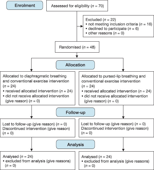
The sample size of 48 stroke patients was estimated using G*Power 3.1.9.4 for Windows. The effect size was 0.39, the confidence interval was 95%, and the power of the study is 90%, considering 10% dropouts. Participants were randomly allocated at a 1:1 ratio by computer-based randomisation into group A (n = 24) that received diaphragmatic breathing and conventional exercises, and group B (n = 24) that received pursed-lip breathing and conventional exercises. Both groups underwent the interventions 5 days per week for 4 weeks, with the interventions starting concurrently for both groups immediately following randomisation to ensure uniformity in the study timeline.
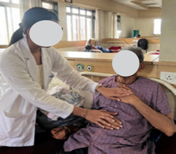
The patients in group A performed diaphragmatic breathing to enhance diaphragm engagement and abdominal breathing control. Patients were positioned in a semi-upright position, with one hand placed on the chest and the other on the abdomen to monitor movement, with the backrest elevated at a 30–45-degree angle. They were instructed to inhale for 3 s deeply through the nose, ensuring visible abdominal expansion while minimising chest movement. The breath was held for 5 s, followed by a slow exhalation through the mouth for 4 s. This cycle was performed at a rate of 6–8 breaths per minute, repeated 10 times per session, for three sessions daily. A one-minute rest period was provided between sessions to prevent fatigue and ensure recovery. The primary goal was to promote effective diaphragmatic movement and reduce accessory muscle use, as supported by literature on respiratory rehabilitation in stroke patients [20]. A demonstration of diaphragmatic breathing is shown in Figure 1.
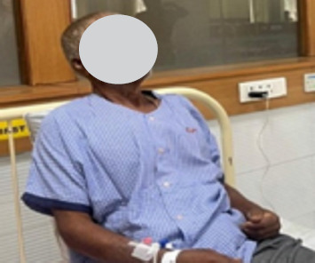
The patients in group B performed pursed-lip breathing to regulate airflow and enhance diaphragmatic activation. Patients were positioned in a semi-upright position, with one hand on the chest and the other on the abdomen to monitor breathing patterns, and with the backrest elevated at a 30– 45-degree angle. They were instructed to inhale for 3 s deeply through the nose, ensuring abdominal expansion, followed by a slow, controlled exhalation through pursed lips (as if whistling) for 6 s to maintain a prolonged exhalation phase. This cycle was performed at a rate of 6–8 breaths per minute, repeated 10 times per session for three sessions daily. A one- minute rest period was provided between sessions to allow for recovery. The goal was to regulate chest and abdominal pressure, improve airflow control, and enhance diaphragmatic engagement, consistent with established physiotherapy protocols [11]. A demonstration of pursed-lip breathing is shown in Figure 2.
Patients were instructed to pause the exercise and resume normal breathing if they experienced hyperventilation symptoms (e.g., dizziness, tingling, or lightheadedness). To ensure proper technique, adherence to the protocol, and patient safety, all the exercises were supervised by trained physiotherapists. Moreover, both groups performed additional conventional exercises in a single daily session, 5 days per week, with each exercise performed for 2 sets of 10 repetitions unless otherwise specified, progressing to 3 sets by week 3 if tolerated. Stretching exercises for the biceps, ham-strings, and adductors were performed in a seated or supine position (depending on patient ability), holding each stretch for 20 s to improve flexibility. Active-assisted range of motion (ROM) exercises included shoulder (0–90°), elbow (0–90°), wrist (0–60°), and hip (0–90°) flexion/extension, hip (0–30°) abduction/adduction, and knee flexion/extension (0–90°), with a physiotherapist or caregiver assisting to achieve full range as needed, performed in supine or seated positions. Thoracic expansion exercises involved deep inspiratory efforts with the hands on the lower ribs to encourage lateral chest expansion, performed for 10 breaths in a semi-upright position. Scapular sets involved 10 repetitions of shoulder blade squeezes in a seated position to strengthen the scapular stabilisers. Trunk rotations were performed seated, with patients rotating their upper body 10 times per side to a comfortable range. Abdominal curls (partial sit-ups) were conducted in a crook lying position, with 10 repetitions to engage core muscles. Pelvic bridging involved lifting the pelvis off the bed in a crook lying position for 10 repetitions, holding each lift for 3 s. Transfers (e.g., bed-to-chair) were practiced 5 times per session, with assistance as needed to improve functional mobility. To prevent fatigue, a 30-second rest was provided between exercises.
Progression was individualised based on patient tolerance if no adverse effects were reported. All exercises were conducted on a treatment bed or chair, with no additional equipment beyond a pillow for head support during the supine exercises. The outcome measures that were assessed at baseline (Day 0) and post-intervention (after four weeks) consisted of the TIS to evaluate trunk control [21–23], and maximum inspiratory and expiratory pressures (measured using a MicroRPM respiratory pressure meter) [17]. The TIS consists of 17 items that use a 2-, 3-, or 4-point ordinal scale for assessment and evaluate trunk coordination and sitting balance, both dynamic and static. The total scores range from 0 to 23, where higher scores indicate better performance. The scale has demonstrated an intraclass correlation coefficient (ICC) of 0.98, reporting high reliability. Administering the TIS typically takes between 6 and 16 minutes. During the assessment, patients received verbal instructions, and demonstrations were provided if needed. Adjustments were made between attempts to ensure accuracy. The test started with the patient seated unsupported on the edge of a bed or treatment table. Their head and body were aligned at the mid-line, thighs supported, knees bent at a 90-degree angle, feet flat on the floor, and arms resting on the legs [21–23].
The MicroRPM is a compact, portable, and non-invasive mouth-pressure manometer equipped with a mouthpiece and a small monitor for displaying results in centimetres of water. Before the actual measurements, each participant completed five maximum inspiratory and five maximum expiratory warm-up attempts to familiarise themselves with the procedure. For MIP and MEP assessments, the participant sealed their lips tightly around the flanged mouthpiece and held the gauge with both hands. To measure MEP, they were instructed to inhale as deeply as possible and then exhale forcefully for more than a second against the gauge’s resistance. Conversely, for MIP, they exhaled fully to the residual volume before inhaling maximally for over a second while the gauge provided resistance [17]. Data were collected at baseline and post-intervention, and all recorded measurements were documented for analysis. Microsoft Excel was used for the initial data entry and organisation, including variables such as age, sex, affected side, MicroRPM values, and TIS scores. Data integrity was ensured through double-entry validation.
Statistical analysis
Data analysis was conducted with IBM SPSS Statistics (Version 23). The Shapiro–Wilk test was used to confirm a non-normal distribution (p < 0.05), prompting non-parametric methods. The Wilcoxon signed-rank test was used to assess within-group changes, while the Mann–Whitney U test was used to evaluate between-group differences, with statistical significance set at p < 0.05 and a 95% confidence interval. Figure 3 illustrates the flow diagram representing the study design.
Results
The baseline demographic characteristics of the stroke patients are presented in Table 1. The analysis indicates that the groups were comparable at baseline, with no significant differences in age, sex distribution, or affected side, ensuring a balanced randomisation process.
Table 1
Baseline demographic data
| Variable | Group A | Group B | p-value |
|---|---|---|---|
| Age (years, mean ± SD) | 60.08 ± 12.13 | 61.64 ± 8.66 | 0.765 |
| Sex (n, male/ female) | 21/3 | 22/2 | 0.30 |
| Side affected (n, left/right) | 13/11 | 8/16 | 1.00 |
The analysis of the TIS, MIP, and MEP of the stroke patients within the respective groups is reported in Table 2. Both groups demonstrated statistically significant improvements in trunk control and respiratory muscle strength from baseline to post-intervention.
Table 2
Within-group analysis of TIS, MIP, and MEP
The analysis of the TIS, MIP, and MEP of the stroke patients between the groups is reported in Table 3. No significant differences were observed between groups at baseline or post-intervention for any of the outcome measures, indicating that the two breathing interventions combined with conventional exercises yielded comparable improvements.
Table 3
Between group analysis of TIS, MIP and MEP
The comparison of TIS, MIP, and MEP according to the effect size is reported in Table 4. The larger effect sizes in group A for all outcomes suggest that diaphragmatic breathing may have a slightly greater clinical impact on trunk control and respiratory muscle strength compared to pursed-lip breathing.
Discussion
This study investigated the comparative effects of diaphragmatic versus pursed-lip breathing, both combined with conventional exercises, on trunk control and respiratory muscle strength in sub-acute stroke patients. Both interventions led to statistically significant within-group improvements after four weeks. However, no significant between-group differences were observed in these outcomes, indicating comparable efficacy between the two breathing techniques. Notably, the effect sizes were consistently higher in the diaphragmatic breathing group across all measures compared to the pursed-lip breathing group, suggesting a potentially greater clinical impact for diaphragmatic breathing despite the lack of statistical superiority.
These findings are supported by several studies highlighting the benefits of respiratory exercises in stroke rehabilitation. For instance, Seo et al. [16] demonstrated that both diaphragmatic and pursed-lip breathing significantly enhanced respiratory muscle activation in chronic stroke patients, with diaphragmatic breathing showing greater effects on inspira-tory muscles, aligning with the higher MIP effect size in the present group A. Similarly, Wu et al. [18], in a systematic review and meta-analysis, found that respiratory muscle training, including diaphragmatic techniques, improves strength and reduces complications in stroke survivors, corroborating the present within-group MIP and MEP outcomes. Song and Park reported that chest expansion exercises similar to diaphragmatic breathing improved TIS scores and trunk control ability in stroke patients, consistent with the TIS improvements in both groups in the current results [24]. Cabanas- Valdés et al. [25] showed that additional core stability exercises enhanced dynamic sitting balance and trunk control in subacute stroke patients, supporting the role of respiratory- linked interventions. Zheng et al. [6] compared Liuzijue Qi-gong (incorporating diaphragmatic elements) to conventional training and observed superior trunk control and respiratory function, reinforcing the trend of larger effect sizes with diaphragmatic approaches. Pai and Li [3], in a scoping review and meta-analysis, noted a positive relationship between trunk control and respiratory function in stroke patients, emphasising how diaphragmatic breathing facilitates trunk-limb coordination through simultaneous diaphragm and abdominal muscle activation.
Moreover, An et al. [26] highlighted immediate benefits of diaphragmatic breathing on pulmonary function via improved diaphragm excursion in chronic stroke patients. Haruyama et al. [27] found that core stability training improved trunk function, standing balance, and mobility in stroke patients, aligning with the trunk control enhancements in the current study. Khallaf [28] reported that task-specific training improved trunk control and balance in subacute stroke patients. Howard et al. [29] underscored the pathophysiological aspects of breathing post-stroke, noting respiratory muscle strength’s role in preventing complications like aspiration pneumonia. Zhang et al. [30], in a meta-analysis, showed that respiratory muscle training enhances function and reduces complications in early stroke, mirroring the overall benefits of the present study. Kong and Krishnan [31] described truncal impairment post-stroke impacting functional outcomes, while Rasheed et al. [2] found diaphragmatic breathing with abdominal drawing-in improved trunk stability in stroke patients.
Contrasting or complementary studies provide nuance to the present non-significant between-group differences and varying respiratory impairments. For example, Shetty et al. [7] compared diaphragmatic breathing to incentive spirometry in stroke subjects and found no significant differences in respiratory function, attributing the similar outcomes to shared diaphragmatic excursion mechanisms, in parallel with the current comparable group effects. Nguyen and Duong [11] described pursed-lip breathing’s role in prolonging expiration and reducing dyspnoea but with limited direct impact on inspiratory strength, consistent with the slightly lower MIP effect in group B in the current study. Breslin et al. [32] found that pursed-lip breathing increased inspiratory accessory muscle recruitment, associated with increased dyspnoea in some COPD contexts, potentially explaining why it may be less effective for inspiratory-focused outcomes in stroke compared to diaphragmatic breathing. In contrast, Bianchi et al. [33] reported that pursed-lip breathing extended expiratory duration and total breathing time, reducing dyspnoea regardless of pulmonary hyperinflation, highlighting its utility for expira-tory control but possibly limiting trunk stability gains, as seen in the effect sizes of the present study. Kubo et al. [34] observed recovery of respiratory muscle strength post-stroke, with MIP and MEP significantly lower at 3 months compared to healthy subjects, suggesting time-dependent improvements that align with the sub-acute phase gains in the present study but no technique superiority. Thijs et al. [8], in a Cochrane review on trunk training after stroke, noted benefits for balance and mobility but limited evidence on respiratory integration. An et al. [26] found immediate effects of diaphragmatic breathing with cervical mobilisation on pulmonary function in chronic stroke, but without direct comparison to pursed-lip breathing.
Clinically, these findings imply that both diaphragmatic and pursed-lip breathing can be integrated into subacute stroke rehabilitation to improve trunk control and respiratory function, with diaphragmatic breathing potentially preferred for patients requiring greater emphasis on inspiratory strength and core stability. This could reduce the risk of complications, such as aspiration pneumonia, and enhance functional independence, informing tailored physiotherapy protocols and warranting larger trials to confirm long-term benefits.
Limitations
First, the small sample size, while calculated to achieve adequate power, may have limited the ability to detect statistically significant between-group differences, particularly given the observed trend of larger effect sizes in the diaphragmatic breathing group. A larger sample could enhance the statistical power to confirm whether diaphragmatic breathing offers superior clinical benefits over pursed-lip breathing. Second, the duration was limited to four weeks, which may not have been sufficient to capture the long-term effects of the interventions.
Conclusions
This randomised controlled trial demonstrated that both diaphragmatic breathing and pursed-lip breathing, when combined with conventional exercises, significantly improved trunk control and respiratory muscle strength in subacute stroke patients over a four-week intervention period. Both interventions yielded comparable outcomes, as evidenced by significant within-group improvements in trunk impairment scale (TIS) scores, maximal inspiratory pressure (MIP), and maximal expiratory pressure (MEP), with no statistically significant between-group differences. However, the consistently higher effect sizes observed in the diaphragmatic breathing group suggest a potential for greater clinical impact, particularly in enhancing inspiratory strength and core stability. These findings underscore the value of incorporating targeted respiratory exercises into stroke rehabilitation protocols to address trunk control and respiratory dysfunction, which are critical for functional recovery and reducing complications such as aspiration pneumonia.