
Introduction
Stroke continues to rank as the second leading cause of death (roughly 7 million) and the third leading cause of death and disability combined (measured by disability-adjusted life-years lost, or DALYs; more than 160 million DALYs) among non-communicable diseases (NCDs), according to the most recent Global Burden of Disease (GBD) 2021 stroke burden estimates. This is the most thorough GBD stroke epidemiology study to date, and it demonstrated that between 1990 and 2021, the number of stroke victims, stroke deaths, and stroke-related disabilities increased significantly worldwide: incident strokes increased by 70% (95% UI 66 to 75), stroke deaths increased by 44% (32 to 56), and DALYs increased by 32% (22 to 43). The majority of the global stroke burden (87.2% of deaths and 89.4% of DALYs) occurred in low-income and low-middle-income countries (LMICs). About 80% of stroke victims experience hemiparesis and spasticity, which are motor dysfunctions that impede their independence and everyday activities [1].
One of the most common long-term effects of stroke is impaired hand function. Up to 87% of stroke patients experience immediate paralysis of the hand or upper limb. Following a stroke, some motor control recovery is normal; this usually peaks during the first three months and reaches a plateau by six months. However, 3–6 months after a stroke, 40–80% of stroke survivors have not fully recovered their upper extremities [2].
Chronic stroke patients frequently experience hand dys-function after primarily ischemic stroke, with both positive and negative symptoms. The former includes flexion synergy and loss of fine hand movements, while the latter includes flexion and hand flexion, which facilitates the patient’s ability to perform daily living activities [3].
Patients who have had a stroke often experience cognitive deficits, sleep issues, anxiety, low self-esteem, and trouble completing activities of daily living [4].
A form of rehabilitation known as tele-rehabilitation intervention uses information and communication technologies (ICT) to deliver rehabilitation services to patients wherever they may be. The patient receives this kind of therapy through a device with an Internet connection, such as a laptop, computer software, smartphone application, or videoconferencing [5]. In addition to helping patients who can visit the clinic, tele-rehabilitation also aids patients with disabilities who are unable to travel to the clinic for rehabilitation services [6].
Through tele-rehabilitation, patients can be exposed to a variety of evaluation and therapy techniques using robotic, virtual reality, and remote monitoring technologies [7]. Both acute and chronic cases of neurological dysfunction are treated in this way [8]. Patients can be exposed to tele-rehabilitation through software, videoconferencing, virtual reality, artificial intelligence, or mobile phone applications. Increasing patient encouragement during the exercises can improve the efficacy of rehabilitation regimens [9]. Therefore, the current study tried to investigate the effect of tele-rehabilitation intervention on hand, cognitive functions and depression of hemiparetic patients. The hypothesis of the study suggested the null hypothesis that there was no statistically significant effect of tele-rehabilitation intervention on hand, cognitive functions and depression of hemiparetic patients.
Subjects and methods
Study design
The current randomised controlled trial double-blinded study was conducted at Cairo University’s Faculty of Physical Therapy’s Neurology Outpatient Clinic as well as each patient’s house.
Participants
Thirty hemiparetic patients were chosen from Cairo University’s Faculty of Physical Therapy’s Neurology Outpatient Clinic. Both sexes of patients were included. The patients were referred by a neurosurgeon or neurologist, and magnetic resonance imaging (MRI) or computed tomography (CT) was used to confirm the diagnosis.
Criteria for inclusion
Patients between the ages of 45 and 55, were included in this study. The patients with a duration of illness between three and six months. According to the modified Ashworth scale (MAS), the patients’ hand spasticity ranged from grade 1 to +1. The patients had a FMUE (Fugl-Meyer upper extremity) score of at least nine, indicating mild hand function. On the Beck Depression Inventory questionnaire, the patients’ scores ranged from 17 to 20 (borderline clinical depression). Every patient had good vision and hearing. The patients successfully filled out the consent form and completed the assigned activities. In order to enable video communication while at home, patients need to have at least ten years of education to be able to use a smart device that is connected to the Internet, such as a laptop, phone, or tablet. In addition to medical care, the patients were instructed to not receive any other kind of treatment. The patients should be able to communicate and obey commands with ease. The patients’ MMSE scores were over 21, indicating good cognitive abilities.
Criteria for exclusion
Not included in this research: Individuals with unstable medical conditions, such as unstable angina, symptomatic heart failure, uncontrolled diabetes mellitus or hypertension. Patients who are unable to utilise the video communication device, have moderate-to-severe impairment of hand functions, or have any other pathology impacting hand function except stroke. Patients who have visual or hearing impairments. Patients who have communication issues or who have trouble understanding or obeying instructions. Individuals who have seizures or psychological disorders. Patients who have not had a stroke but have another illness that impairs cognitive function. Individuals who have a history of stroke or other neurological conditions.
Sample size determination
The G*POWER statistical analysis software (G*power version 3.1) is used to determine the necessary sample size a priori for study groups. A two-sided t test with an alpha level of 0.05, a power of 95%, and an effect size of 1.4 was calculated based on the results of a pilot study involving 15 patients, yielding a sample size of 15 patients per group.
Randomisation and concealed allocation
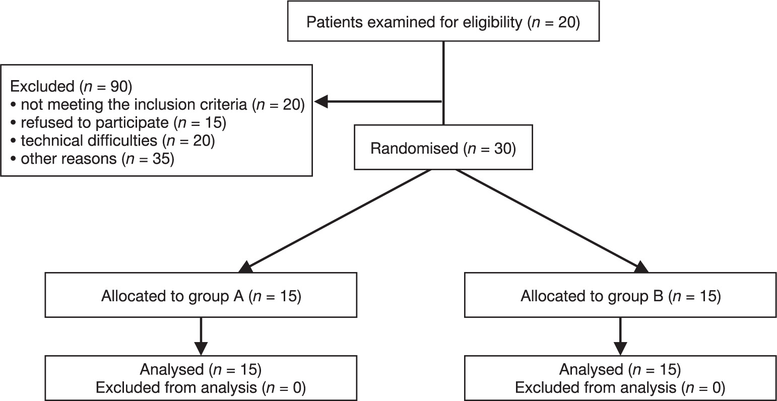
Among the eligible individuals, two groups were chosen at random: group A and B. The allocation was carried out by a researcher who was blind to the recruiting and treatment of the participants. The consort flow diagram with the hemiparetic patients included is shown in Figure 1. A numeric table that was produced at random was used to conceal the allocation. The intervention groups were recorded on individually numbered index cards arranged in a sequential fashion. The cards were then folded and placed inside opaque, sealed envelopes. The assignment group claims that on the day of the initial assessment (assessor blinding), a different therapist who was not informed of the results of the initial assessment opened the envelope and started the treatment. Patients were not aware of their assignment group and the intervention by the other group. In order to teach patients (patient blinding) in the tele-rehabilitation group how to complete exer- cises at home and to determine when it is appropriate for patients to complete sessions at home, the first session of the treatment program is implemented at the clinic for both groups.
Procedure
The 30 hemiparetic patients were divided into two equal groups at random: the experimental group (GA) (n = 15) received the hand rehabilitation program through the tele-rehabilitation program, a technological device, and the control group (GB) (n = 15) received the same hand rehabilitation program at the physical therapy clinic. Patients in both groups underwent a face-to-face examination at the clinic prior to the commencement of their rehabilitation program, and they underwent a final examination. The rehabilitation was conducted three times a week for a total of twelve weeks (36 sessions).
The control group (GB) received a hand rehabilitation program at the rehabilitation centre, while the experimental group (GA) received the rehabilitation program via laptops equipped with a camera, microphone, and a reliable Internet connection for the upcoming sessions.
Interventions
Patients in the tele-rehabilitation group are contacted via laptops that have cameras, microphones, and a reliable Internet connection. The therapist’s laptop camera is positioned so the patient may watch the therapist while the therapist demonstrates the exercises. The patients’ laptop webcams were also set up so the therapist could watch them as they completed the exercises. From among the exercises covered by neurodevelopmental treatment, those that were remotely applicable and suitable for the patients were selected. After assessing each patient, the exercise plans were developed by selecting suitable exercises from the list below that were pertinent to the needs and requirements of the individual. The number of exercise repetitions was calculated based on each person’s requirements and preferences. The activities were selected to be appropriate for the group of tele-rehabilitation patients to perform at home. The following exercises are part of the workout program:
The hand rehabilitation program is used for patients in both groups; for the GA, it is used on the patient’s computer at home, while for the GB, it is used at the clinic. However, the initial session is used at the clinic for both groups to show the patient how to perform the exercises. The following exercises are part of the program: For the GA: After the therapist performs the exercise on himself, the patient must see the therapist via a laptop camera and then perform the same activity again. For the GB, the therapist demonstrates the movement with active aided movement before the patient completes it on their own.
The hemiparetic patients in both groups performed the following exercises:
A. Free exercises
Wrist flexion and extension: The patient places their arm on the table’s edge, palm down, then raises it, repeating with the palm up. This is done while they are comfortably seated.
Wrist side movement: The patient is instructed to place their palm down on the table and then move their wrist side to side.
Thumb flexion and extension: The patient sits comfortably, opens their hand to indicate the number five, and then flexes their thumb by moving it toward their little finger to indicate the number four. This process is repeated.
Finger opposition: The patient is instructed to flex the affected arm by placing the elbow on the table, making a ring with the index and thumb tips, then repeating the motion with the middle, ring, and pinkie fingers before letting go. This is done twice for each finger.
Palm up and down: While seated, the patient places one hand on the table with the palm facing up and free from the table. Then, using the other unaffected hand, the patient moves the palm down, then up, and so on, repeating the motion up to ten times. The mobility of the hands and fingers is improved by this workout.
B. Ball exercises
Ball grip: The patient is instructed to squeeze the ball tightly in their hand, hold it, and then release it. This exercise is repeated ten times for two sets.
Finger flexion: The patient is instructed to hold the ball in their palm and press the fingers into the ball while flexing them. The focus is on the ball’s overall global grasp movement.
Thumb extension: The patient is instructed to place the ball between the flexed thumb and the two fingers that are outstretched. The patient is then instructed to roll the ball while performing the exercise ten times for two sets.
The pinch exercise involves having the patient place the ball between their thumb, index, and middle finger, squeeze, hold, and then release. They are to perform this exercise ten times for two sets.
Opposition: The patient is told to bring their thumb to their little finger while holding the ball in the palm of their hand. Over the course of two sets, they must do this activity ten times.
Side squeeze: The patient is asked to put the ball between 2 fingers while squeezing it, hold then relax, repeating the exercise 10 times for 2 sets.
Extend out: The patient is instructed to place the ball on the table, place the tips of their fingers on it, and roll it outward. This exercise is repeated ten times for two sets.
C. Household exercises
Roll movement: The patient is instructed to place their affected arm on the table, place a bottle of water in the affected hand, and then, while keeping the bottle in a relaxed position, curl the fingers and release the bottle. This exercise is repeated ten times for two sets.
Wrist curl: The patient is instructed to grasp a water bottle in the affected hand while holding it up with the unaffected hand. This allows the wrist to extend and curl downward, then upward. The exercise is repeated ten times for two sets.
Extending the wrist: The patient is instructed to grasp a water bottle in the affected hand while holding it up with the non-affected hand. The wrist should be extended, and the exercise should be repeated ten times for two sets.
D. Pen exercises
Pinch and release: The patient is instructed to place a pen on the table, grab it with the afflicted fingers, slide it across the table, and then let it go. This exercise is repeated ten times for two sets.
Pen spine: To isolate thumb and finger movements, the patient is instructed to place the pen on the table and use their thumb and fingers to spin it as quickly as possible for 15 s without moving the shoulder.
E. Exercises with coins
Coin drop: The patient is instructed to place eight consecutive quarters in the palm of the afflicted hand, then slide one quarter down to the thumb and index finger with the thumb, pinch it with the thumb and index fingers, and set it on the table while holding the other quarters in the hand. Finally, the patient is instructed to repeat the process with the remaining quarters.
Coin stacking: In this exercise, a variety of coins of varying sizes were provided, and the patient was instructed to arrange them according to size, placing the larger coins lower and the smaller coins higher.
Outcome measurement
The experimental and control groups were assessed before and after a 12-week treatment period using the nine-hole peg test, FMUE, MOCA (Montreal cognitive assessment scale), REHACOM system (attention concentration, and figural memory), and Beck Depression Inventory questionnaire.
1. The Nine Hole Peg Test: This is a tool used to evaluate hemiparetic patients’ finger dexterity. It was carried out in accordance with earlier instructions [10].
Goal: This exam evaluates finger dexterity and is regarded as a therapeutically valuable way to gauge upper limb function.
Description and equipment used: A wooden board with nine holes that are 10 mm in diameter and 15 mm deep, spaced 32 or 50 mm apart, is used for the nine-hole peg test. Beside the board is a shallow, dish-shaped container for the pegs. The nine pegs measure 32 mm in length and 7 mm in diameter. A stopwatch is also required [11].
Procedure for the nine-hole peg test: The patient is asked to use only the hand being assessed to swiftly insert each peg into the holes on the board after taking them out of a container. After that, the patient is told to take each peg out of its hole and put it back in the container. When the patient touches the first peg, the therapist starts the stopwatch, and when the last peg is in the container, the stopwatch is stopped. The stopwatch is used to record the overall amount of time needed to finish the task. The nondominant hand is used for two consecutive trials after two consecutive trials with the dominant hand [12].
Scoring: The mean time was calculated by repeating the experiment three times, and the time needed for patients to insert the nine wooden sticks into the holes was noted (this method was employed in our study assessment). Counting the number of pegs positioned in 50 or 100 s is an additional score option. In this instance, the number of pegs set per second is used to describe the results [13].
2. FMUE: This specific test is intended to assess the motor functioning and impairment of stroke patients. An ordinal scale is used to assess 33 items for upper extremity function, with a maximum score of 66 [14].
Following a stroke, this scale is used to quantify motor disability. The clinically significant difference for determining the improvement in upper extremity function in the FMUE is approximately a six-score change. The FMUE manual (version 7) was developed by a team at the University of California, Irvine, under the direction of Dr Steven Cramer. This scale is intended to evaluate motor functioning, balance, joint functionality, and sensation in people who have suffered a stroke [15].
FMUE procedure: This exam evaluates motor skills such as movement, coordination, and reflex actions of the hand, wrist, forearm, elbow, and shoulder. These items evaluate upper extremity recovery and incorporate Brunstrom’s stages of motor recovery [14].
A bed or mat is needed for this test, and several tools and small objects – such as paper, a ball, cotton ball pencil, reflex hammer, cylinder, goniometer, stopwatch, blindfold, chair, and bedside table – are used to evaluate motor function, sensation, reflexes, and range of motion. When the patient is in a comfortable resting posture, the therapist employs these tools to assess the patient and watch what they do.
Regarding FMUE scoring, it is based on the patient’s performance and their ability to finish the task using a 3-point rating system: 0 for not being able to complete the task, 1 for partially completing it, and 2 for finishing it [16].
3. MOCA: This quick screening exam is intended to identify moderate cognitive impairment (MCI) and assess the various cognitive functions of stroke patients. It takes roughly ten minutes to complete and has thirty points. Visuospatial skills, executive functions, short-term memory recall, attention concentration, working memory, language, and orientation to time and space are among the cognitive abilities evaluated in the MOCA [17].
The MOCA procedure involves giving the stroke patient a sheet of the MOCA scale and having them sit comfortably. The MOCA has 30 questions and takes roughly 10 minutes to finish. The MOCA cutoff score is 26, thus subjects with a score of 26 or higher are regarded as normal [18]. The total score is 30 points.
4. REHACOM system: The REHACOM system was made by Hasomed REHACOM (CE certified, EN-ISO-13485), Schuh frted (model No. 454, D-14482 posted am, Karl-Liepknecht, Austria), and G S: REHACOM Version 5. Basic Manual. 2003, 2012. It is a software program made for diagnosing and treating patients with cognitive impairments. Figural memory (FM), attention concentration (AC), reaction behaviour (RB), and logical reasoning (LR) are their four categories. Both the attention concentration and figural memory categories were evaluated for the hemiparetic individuals in this study.
Procedure: After entering each stroke patient’s data, the therapist configures the REHACOM system’s settings based on the category to be utilised for the evaluation or treatment. Attention concentration is the category employed in this investigation. The evaluation lasts for thirty minutes. The patient is seated next to the therapist, who initially explains how to operate the REHACOM equipment and its panel.
After learning how to operate the system, the patient settles comfortably at the level of the REHACOM screen. The patient takes the test both before and after the rehabilitation program is over, and the system generates a report at the conclusion of the program.
Regarding the attention concentration category, there are 24 levels of difficulty, a distinct image that is compared to a matrix of various images that are displayed in front of the patient, and only one image that is exactly like it. As the levels advance, the difficulty increases. A report with the following information is generated at the conclusion of the test: difficulty level, maximum reaction time, and minimum reaction time.
In order to measure figural memory, the picture-to-picture method was employed. There are nine different levels of difficulty. It is divided into two stages: the acquisition phase, where a predetermined number of images are shown to the patient, and the reproduction phase, where a series of images, including the previously displayed images, scroll past and the patient must identify the previously displayed images. Following the test, a report is produced that includes the following information: acquisition time, solution time, and difficulty level.
5. BDI. This questionnaire serves as both a screening tool for depression and a means of determining the severity of depression [19]. It has 21 elements, each of which represents a distinct depressive symptom. Physical or somatic, affective or emotional, cognitive, and vegetative symptoms, including eating and sleep patterns, are the four categories of items that were evaluated [20].
The questionnaire’s overall score ranges from 1 to more than 40. A score of 1 to 10 is regarded as normal, a score of 11 to 16 as mild mood disturbance, a score of 17 to 20 as borderline clinical depression, a score of 21 to 30 as moderate depression, a score of 31 to 40 as severe depression, and a score of more than 40 as severe depression. A higher questionnaire score therefore indicates a more severe case of depression in the patient [21].
The BDI procedure is as follows: After settling into a comfortable sitting posture, the patient is asked to rate the 21 depression items on a scale of 0 to 3 to indicate how depressed they are. The patient is then given the score [22].
Statistical analysis
IBM Corp., Chicago, USA, 2013’s statistical program for social sciences (SPSS) version 22 was used to gather and analyse the data for this study. The mean ± standard deviation (SD) was used to express the quantitative data. Frequencies and percentages were used to express the qualitative data. In the quantitative data, two independent variables were compared using the independent t test, and two dependent variables were compared using the paired t test, within-group comparisons were analysed using the Wilcoxon signed-rank test, and between-group comparisons using the Mann–Whitney U test. A p-value of less than 0.05 was deemed significant, while a p-value of less than 0.01 was deemed extremely significant.
Results
This study looked at how tele-rehabilitation programs affected the depression well-being, hand and cognitive abilities of hemiparetic patients. This study involved 30 hemiparetic patients who were split into two equal groups at random: the experimental group (GA) and the control group (GB). The MOCA, the REHACOM system, FMUE, the nine-hole peg test (9HP), and the BDI were used to evaluate both groups before and after a 12-week (36-session) rehabilitation program.
Subject characteristics
Thirty hemiparetic patients in all were included in this study, with fifteen patients in each group. The consort flow diagram with the hemiparetic patients included is shown in Figure 1. The mean age of the patients was 49.267 for the GB and 50.6 for the GA, with an age of 45 ± 55 years. Table 1 displays the groups’ general characteristics.
Table 1
Demographic data
According to the results of the Nine-hole Peg Test (9HPT), the GB undergoing rehabilitation and the GA undergoing tele-rehabilitation did not differ statistically significantly (p-value = 0.3332, as illustrated in Table 2).
Table 2
Mean values of 9HT
Regarding FMUE, the study’s findings demonstrated a statistically non-significant difference (p-value = 0.2450) between the experimental groups that received the tele-rehabilitation system and the control group after completing the rehabilitation program (Table 3).
Table 3
Mean values of FMUE
Regarding the REHACOM system’s attention concentration level, the results indicated that there was a highly significant difference (p-value = 0.0001) between the experimental groups that received the tele-rehabilitation program and the control group after receiving the program, favouring the GA who received the tele-rehabilitation program (Table 4).
Table 4
Mean values of attention concentration level
The results indicated that there was a highly statistically significant difference (p-value = 0.0001) between the experimental groups receiving the tele-rehabilitation program and the control group after receiving the rehabilitation program with regard to the figural memory level of the REHACOM system, favouring the GA receiving the tele-rehabilitation program (Table 5).
Table 5
Mean values of figural memory level
According to the MOCA results, there was a statistically significant difference between the experimental group that received the tele-rehabilitation program and the control group, favouring the GA. The experimental group A’s p-value was 0.0001 before and after the rehabilitation program, and the groups’ p-values were 0.0019 and 0.0019, respectively (Table 6).
Table 6
Mean values of MOCA scale
Regarding the Inventory of Beck Depression, the results of the study demonstrated that there was a highly significant difference between the experimental group receiving the program and the control group after receiving the rehabilitation program in favour of the GA receiving the tele-rehabilitation program (p-value = 0.0001) (Table 7).
Discussion
The purpose of this study was to find out how a tele-rehabilitation program affected the depression and hand and cognitive abilities of hemiparetic patients. In this study, thirty hemiparetic patients were divided into two groups: fifteen in the control group and fifteen in the tele-rehabilitation group.
The results indicated that while there was no significant difference in the nine-hole peg test (9HPT) results, there was a significant difference in the FMUE, cognitive functions, and Beck’s Depression Inventory questionnaire in favour of the GA enrolled in the tele-rehabilitation program.
Since the patient receives the rehabilitation at home rather than in a clinic, it has been discovered that tele-rehabilitation is an inexpensive kind of rehabilitation that also helps the patient’s psychological state. Additionally, the tele-rehabilitation program is only appropriate for patients with mild cases; individuals with severe cases require extra attention and support.
The availability of a good Internet connection is one of the challenges we face while using tele-rehabilitation because, in most cases, the Internet connection is unstable, as is the computer device that the patient uses to receive treatment.
The current study’s findings on the nine-hole peg test indicated that there was no discernible difference between the experimental and control groups. The current study’s 9-hole peg test results are in line with a 2023 study by Alejandro et al. [23], which found that tele-rehabilitation programs were more successful than traditional rehabilitation through clinic appointments and also helped people visit rehabilitation clinics less frequently.
The results of the Fugl-Meyer upper extremities assessment revealed a significant difference between the experimental and control groups in terms of upper extremity function following the use of the tele-rehabilitation system, as opposed to the GA utilising the same equipment.
These findings are in line with those of a study conducted in 2022 by Moulaei et al. [24], who demonstrated that employing the tele-rehabilitation system improved upper extremity function as measured by Fugl-Meyer.
It was discovered that using the tele-rehabilitation system has numerous advantages, such as improving the musculo-skeletal system’s functions, increasing the patient’s motivation and enthusiasm to perform exercises instead of doing them the old-fashioned way, which increases the patient’s likelihood to perform rehabilitation exercises and, consequently, increases patient participation in the rehabilitation program.
The MOCA results indicated that, in favour of the GA, there was a statistically significant difference between the experimental group that received the tele-rehabilitation program and the control group.
Regarding the REHACOM system results, the figural memory and attention concentration levels of the experimental groups that received the tele-rehabilitation program and the control group showed a statistically significant difference favouring the GA. This suggests that tele-rehabilitation programs play a significant role in enhancing the cognitive abilities of hemiparetic patients.
Regarding the enhancement of cognitive functions (figural memory and attention concentration) after completing a tele-rehabilitation program, this can be explained by the fact that while the therapist demonstrates the movement in front of the patient and can show them pictures of the movement, tele-rehabilitation also improves the patient’s figural memory and attention concentration.
The results of this study are in conflict with those of a 2017 study by Hüzmeli et al. [25], which found that tele-rehabilitation programs had no effect on patients’ mental health after completion.
According to a different study by Peretti et al. from 2017 [26], tele-rehabilitation has been used in various medical specialties and has been demonstrated to improve many patients’ physical and mental health as well as their pain levels.
This study’s findings were supported by another study by Lin et al. [27] in 2023, which found that tele-rehabilitation or online activities significantly enhance mental and physical functioning.
As for the Beck Depression Inventory questionnaire results, there was a notable difference between the two groups’ results, favouring the GA receiving the tele-rehabilitation program because receiving tele-rehabilitation outside of the clinic improves the patient’s psychological state and thereby lowers depression.
It was discovered that while the patient receives treatment outside of the clinic, the tele-rehabilitation program helps to enhance their psychological state. It is a good way for the patient to get their therapy without being infected in the age of spreading diseases.
The results of Medina et al. [28], who discovered that tele- rehabilitation increases the effectiveness of rehabilitation, corroborated the findings of the current investigation. Additionally, tele-rehabilitation helps patients feel more encouraged, satisfied, confident, and in a better psychological state.
The current study has some limitations: the small sample size used and the lack of follow-ups for the patients to determine the long-term effect of a tele-rehabilitation intervention on the hand and cognitive functions and depression, which is a must for future research.
Conclusions
Based on the data gathered from this study, it can be said that utilising a tele-rehabilitation program in a particular hand rehabilitation program for hemiparetic patients significantly improved their hand function, cognitive abilities (such as visual memory and attention concentration), and psychological well-being status, all of which improved their functional outcome.